
Skin Tissue Model “SCAR-In-A-Dish”
Challenge
Due to limited regenerative potential, tissue injury in higher vertebrates results in the formation of fibrotic tissue that replaces the normal functional tissue of an organ. Skin scars are formed during the healing process of damaged skin caused by disease or injuries. They can range from normal thin scars, to atrophic, hypertrophic, keloid scars, or striae. Dermal scars present an enormous clinical and cosmetic problem, and affect around 100 million people each year in the western world. Scar formation is a complex multifactorial process with pathophysiologic mechanisms and potential targets still unknown. Thus, current therapies focus mainly on scar management while there is a lack of treatment options directed towards prevention of scar formation. The search for new therapeutics is hampered by the lack of appropriate models that offer further insight into the dynamics of scaring in vivo. Therefore, a reliable assay system that offers a basis for compound screening approaches is urgently needed.
Scar development within 5 days
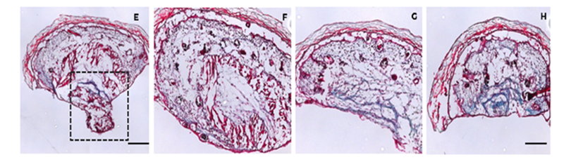
G) and H) Untreated tissue sections (control); E) and F) Tissue sections of SCAD samples treated with Nefopam hydrochloride. The box in (E) marks the reduced scar development under chemical influence.
Technology
The technology offers a novel in vitro model for analysis of scar formation. Skin samples that comprise a full thickness skin sample (epidermis, dermis and subcutis) are cultured up to 5 days untethered in liquid culture. Assay conditions have been developed for 96 and 384-well formats and match the complexity of tissue in vivo by recapitulating the scaring process regarding fiber alignment and architecture of the extracellular matrix, as well as the migration of keratinocytes. The SCAD assay (Scar-in-a-Dish) has been successfully applied to initial screenings of chemical libraries for compounds that reduce or even abolish scar formation within the 2 mm tissue sections. Potential readouts include measurements of scar areas in bright field whole-mount images or by immune histochemical staining of sections, complexity analysis of scar tissue in histochemical staining based on an open-source software, as well as measurements of the tissue elasticity.
Commercial Opportunity
The technology is available for in-licensing or further co-development.
Development Status
Proof of concept experiments were performed with murine and human skin samples and have demonstrated applicability of the assay system for validation of promising novel drug candidates. Positive hits have been confirmed in a mouse model of fibrosis formation.
Patent Situation
A priority claiming patent application has been filed in 2018 followed by a PCT application (WO2019154963 A1). National phases were entered in US, EP and CN.
Further Reading
Dongsheng Jiang, Donovan Correa-Gallegos, Simon Christ, Ania Stefanska, Juan Liu, Pushkar Ramesh, Vijayanand Rajendran, Martina M. De Santis, Darcy E. Wagner, Yuval Rinkevich. Two succeeding fibroblastic lineages drive dermal development and the transition from regeneration to scarring. Nature Cell Biology (2018) Vol 20, p.422–431.

Institute of Origin