
Novel therapy approach for treatment of lung fibrosis
Keywords
Fibrosis, IPF, extracellular matrix, mesotheilial cells, matrix movement inhibition, intrapleural injection, gene therapy, cathepsin B, cystatin A
Invention Novelty
This novel therapy approach is based on the findings that fascia and the transfer of pre-existing extracellular matrix (ECM) plays a crucial role in tissue scarring and organ fibrosis. Physical translocation of pre-existing matrix elements to areas of injury were identified as a novel molecular mechanism for fibrosis initiation. In particular, it was demonstrated that mesothelial cells are causative for such ECM movement. By targeting said mesothelial cells, which form the surface cell layer as the outermost lining of each internal organ, and inhibiting movement of pre-existing ECM towards a site of organ injury, fibrosis initiation and progression could be efficiently prevented in animal models and ex-vivo human samples. In chronic models of fibrosis, tissue regeneration was observed.
For this purpose, cystatin A, a natural inhibitor of the thiol proteases cathepsin B, H and L, was selected for a gene therapy approach to directly address the pleural surface of the lung, to inhibit mesothelial cathepsin B which releases pleural matrix and aggravates pulmonary fibrosis.
Value Proposition
Due to a high level of complexity and patho-mechanisms that are not yet understood, there are currently hardly valid approaches to specifically investigate the molecular processes that lead to the development of fibrosis or to enable the development of drugs against fibrotic changes in various organs. The associated forms of the disease are therefore only treatable to a limited extent and represent a significant health problem.
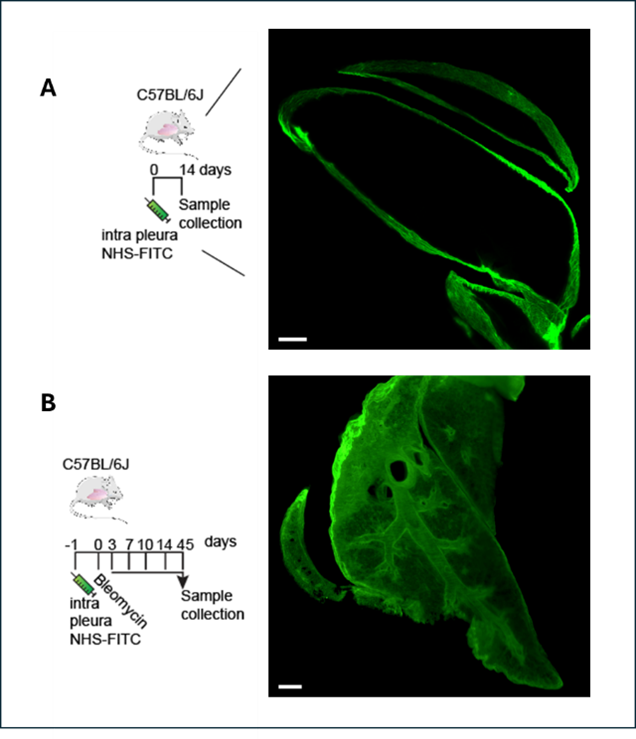
A: Intrapleural administration of a fluorescence label (NHS-FITC) for labeling the ECM of the lung and analysis of the surface ECM labeling after 14 days.
B: After an intrapleural administration of the molecular fluorescence label (NHS-FITC) bleomycin is injected. 45 days after lung injury, a massive transport of the labeled preexisting extracellular matrix into the organ tissue is observed initiating fibrosis.
Technology Description
Cystatin A, a natural inhibitor of the thiol proteases cathepsin B, H and L, was selected for a gene therapy approach to directly address the pleural surface of the lung, to inhibit mesothelial cathepsin B which releases pleural matrix and aggravates pulmonary fibrosis. Intrapleural injections enable direct manipulation of mesothelial cells facing the pleural cavity. As vehicles AAV vectors of the AAV2/8 serotype were applied intrapleurally loaded with therapeutic target gene. In mice, an efficient blockade of the release of fluid matrix components after tissue damage by bleomycin was achieved which prevented ECM movement and fibrotic tissue generation. In chronic models of fibrosis, a regeneration of the fibrotic tissue was observed.
Analysis was performed by labelling preexisting extracellular matrix (ECM) on the surface of different internal organs with N-Hydroxysuccinimide-esters in chemical and viral models of fibrosis, as well as ex-vivo human samples. Cathepsin-mediated cleavage of ECM components was identified as crucial step for mobilization of pre-existing ECM into lung tissue (Fig. B).
Commercial Opportunity
The anti-fibrotic therapy approach, ECM movement modulation as a novel treatment paradigm and ECM labeling assays, are available for in-licensing.
Patent Situation
WO2023/079127A1 has been nationalized in US, CA, EP, CN. IP portfolio on ECM movement modulation and in vitro and in vivo assay systems (WO2020/157122A1; WO2021/028596).
Further Reading
- Fischer et al., Nature Communications 2025; doi: 10.1038/s41467-024-55596-x
- Correa-Gallegos et al., Nature 2019; doi:10.1038/s41586-019-1794-y
- Jiang et al., Nature Communications 2020; doi:10.1038/s41467-020-19425-1
- Wan et al., Matrix Biology 2021; doi:10.1016/j.matbio.2021.01.005
- Fischer et al., Nature Immunology 2022; doi:10.1038/s41590-022-01166-6
- Fischer et al., Nature Protocols 2023; doi:10.1038/s41596-023-00867

Institute of Origin