Efficient multiplex cryo-embedding method for cost-efficient, high-throughput tissue analysis
Keywords
multiplex cryo-embedding & IHC, high-throughput cryo-sectioning, heterogeneous tissues, spatial omics, automated staining, digital pathology
Invention Novelty
The analysis of tissue samples is fundamental to biomedical research and clinical diagnostics. Conventional workflows involve fixation, embedding (e.g., OCT or paraffin), sectioning, staining, and imaging. While reliable, they are labor-intensive, costly, and difficult to scale, especially for parallel processing of multiple, heterogeneous samples. Existing high-throughput (HT) solutions like tissue microarrays (TMAs) are limited by manual processing, repeated freeze/thaw cycles, and constraints on tissue size and shape, increasing the risk of sample damage and variability. The invention introduces a multiplexed cryo-embedding technique using reusable cryo-molds, enabling parallel processing of multiple heterogenous tissues within a single OCT block. The approach allows true HT cryo-sectioning, significantly reducing costs and hands-on time while improving scalability and consistency.
Value Proposition
The method delivers a transformative increase in efficiency, achieving up to 95% reductions in consumable costs (OCT, antibodies, buffers) and hands-on time compared to conventional single-sample workflows or TMAs. By eliminating repeated freeze/thaw cycles, it preserves tissue integrity, morphology, and antigenicity, while avoiding common TMA-related artifacts. Throughput is dramatically increased, as multiple heterogeneous tissues can be embedded and sectioned together in a single block. Slide-to-slide variability is significantly reduced, which is essential for quantitative and multiplex IHC, as well as spatial omics analyses. The method is broadly applicable across clinical diagnostics, research, and pharmaceutical development, particularly where cost, throughput, and reproducibility are limiting factors. By processing all tissues in a single multiplex block, antibody and staining solutions are applied once across all samples, making multiplex panels economically feasible at scale.
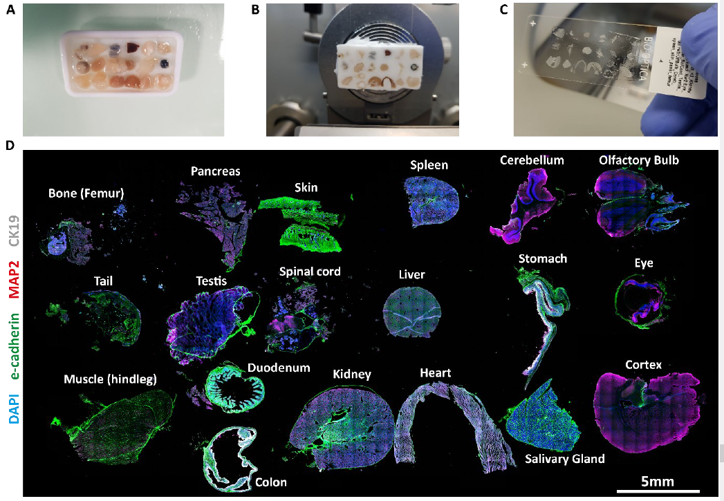
Efficient multiplexed cryo-sectioning. A-C, steps of tissue embedding in a 6x3 grid. D, whole-slide stitched microscopy image of immunostaining for e-cadherin, MAP2, CK19 and DAPI. Individual tissues are labeled within the figure (Reumann et al., unpublished).
Technology Description
The technology uses reusable multiplex tissue molds (MTMs) and a streamlined cryo-embedding procedure. Made from low thermal conductivity, anti-adhesive materials, the molds are durable and allow easy demolding. Each mold has the footprint of a standard microscopy slide and contains recesses to hold individual samples in defined positions. In the workflow, samples are fixed, cryo-protected, and pre-treated with OCT, then placed into the pre-cooled mold, filled with OCT, covered, and placed on dry ice. Once frozen, the OCT/tissue block is removed, inverted so tissues are on top, and an extra OCT layer is added to create a flat cutting surface. The block is then trimmed and sectioned with a standard cryostat. Embedding and freezing all specimens at once produces large, stable sections containing a full panel of tissues, enabling bulk IHC/IF staining and imaging. The workflow is compatible with existing cryostats, adhesion slides, automated stainers, and imaging systems. Although optimized for cryo-embedding, the invention can be analogously applied to paraffin embedding, broadening its applicability,
Commercial Opportunity
The method is especially attractive for providers of cryostats and tissue processing instruments, manufacturers of automated IHC or multiplex staining platforms, and vendors of spatial omics or digital pathology solutions seeking to enhance their offerings. By integrating the technology, companies can upgrade existing platforms with HT cryo-embedding, deliver substantial cost and time savings to pathology labs and pharmaceutical clients, and differentiate themselves through improved reproducibility and scalability in tissue analysis. The technology is available for in-licensing and OEM integration.
Development Status
The innovation has been implemented by multiple labs at IMBA. It has been comprehensively validated, demonstrating robust performance across diverse tissue types, sizes, and stiffnesses, including panels of adult mouse organs (see Figure) and cerebral organoids.
Patent Situation
International PCT application WO2026/022339 A1 has been filed..
Further Reading
Reumann et al. (2025) Cell Reports Methods 5:101023.
Licensing Contact

Dr Torsten Stachelhaus
Technology ManagerT: +49 40 1888 43 48
stachelhaus@ascenion.de
Institute of Origin