
Method and arrangement for identification and correction of optical aberrations
Keywords
aberration corrections, nonlinear microscope, microscopy, biology, life sciences, biomedical research
Invention Novelty
The invention introduces a novel method and arrangement for identifying and correcting optical aberrations, which significantly enhances the resolution and imaging depth capabilities of optical systems like confocal and two-photon microscopes. This method utilizes a dual-beam approach where the intensity of one optical beam is substantially higher than the other, allowing for precise aberration identification through the superposition effects of the two beams. By implementing phase, spatial, and temporal shifts between the beams, the method can detect and analyse radiation excited in the medium to determine aberrations accurately.
Value Proposition
Optical microscopy is an indispensable tool for biomedical research. Yet, the same structures that make biological samples interesting to study under a microscope (such as cells, vasculature, and subcellular organelles) scatter light and thus render tissues opaque. Tissue opacity poses a major challenge to all optical imaging and photo-stimulation methods, fundamentally limiting them to thin sections, cultured cells or superficial layers of tissue. A variety of methods for aberration correction in inhomogeneous media are known. However, these methods are rather slow and their applicability for practical biomedical imaging is limited. The objective of this innovative technology is to facilitate the implementation of aberration corrections in imaging systems.
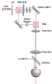
Implementation of aberration correction within a nonlinear microscopy setup. (Source: Papadopoulos et al. 2016)
Technology Description
The innovative method allows the identification and correction of optical aberrations. Thereto a nonlinear microscope is combined with an interferometric unit where the excitation laser beam is split into two arms: one arm acts as the conventional scanning beam that generates an image, the other beam is parked onto the sample. The two beams have significantly different intensities so that the scanning beam acts as a delta like function that, due to interference with the stationary beam, probes the field of the stationary beam. The signal acquired on the photodetector can be shown to be proportional to the field of the aberrated point spread function (PSF) inside the inhomogeneous medium. The knowledge of the amplitude and phase of the light distribution inside the biological tissue allows the compensation of both optical aberrations and tissue-induced scattering.
Commercial Opportunity
The technology is available for in-licensing.
Development Status
The proof of concept has been demonstrated.
Patent Situation
A European priority claiming patent application was filed in 2016 (EP3232245A1), followed by an international PCT-application and granted US-patent (US10962752B2).
Further Reading
Papadopoulos, I. N., Jouhanneau, J.-S. B., Poulet, J. F. A. & Judkewitz, B. Scattering compensation by focus scanning holographic aberration probing (F-SHARP). Nature Photon. 11, 116-123 (2016).

Institute of Origin